Heidelberg University researchers have mapped the cerebellum’s development in humans, mice, and opossums, uncovering its complex structure and significant role in human cognitive evolution. Their findings offer insights into brain development and diseases, with a focus on Purkinje cells and genetic variations over 160 million years.
Scientists at Heidelberg have revealed genetic mechanisms that govern the formation of varied cell types in the human cerebellum and in other mammals.
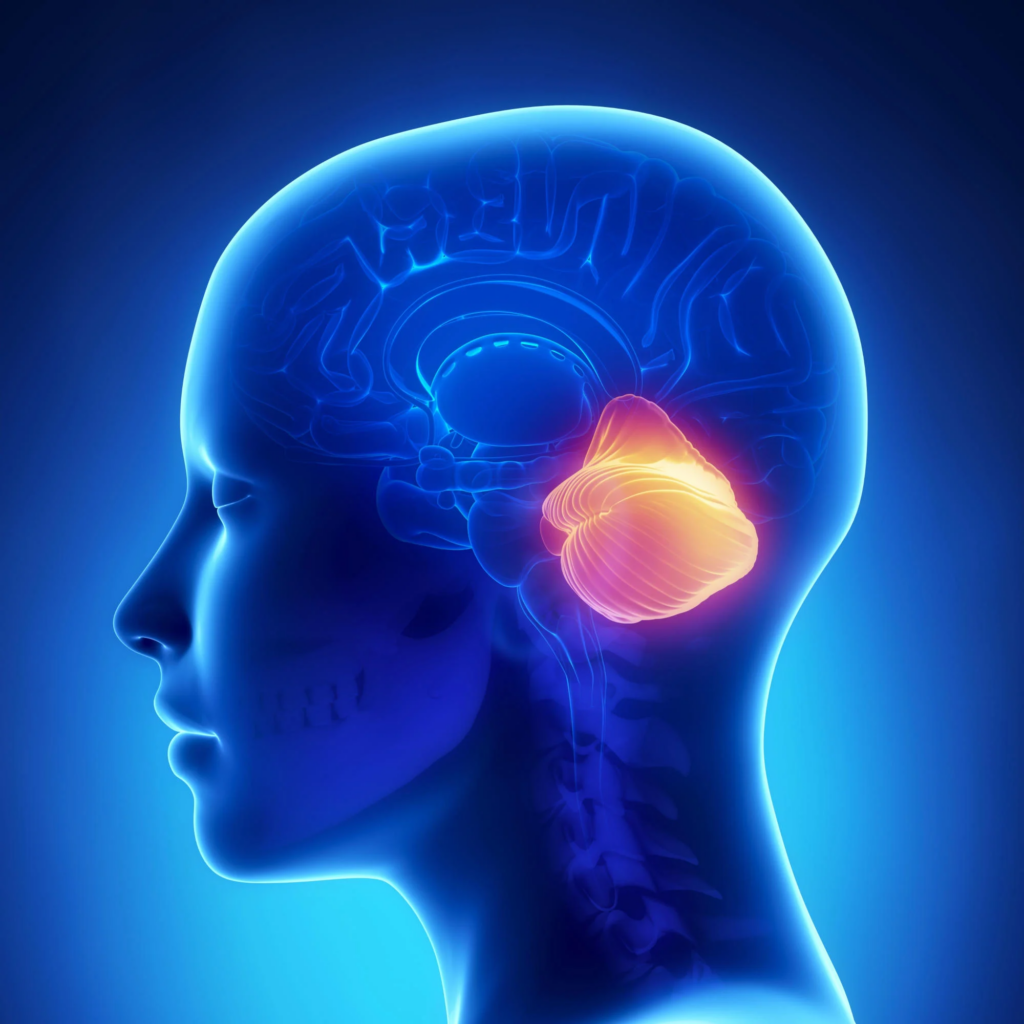
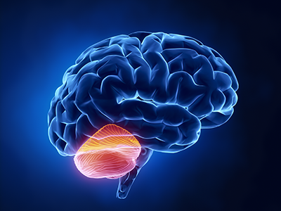
The advancement of higher cognitive abilities in humans is predominantly associated with the growth of the neocortex, a brain area key to conscious thinking, movement, and sensory perception. Researchers are increasingly realizing, however, that the “little brain” or cerebellum also expanded during evolution and probably contributes to the capacities unique to humans, explains Prof. Henrik Kaessmann from the Center for Molecular Biology of Heidelberg University.
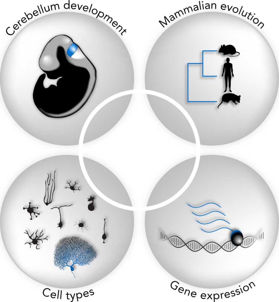
His research team has – together with Prof. Dr Stefan Pfister from the Hopp Children’s Cancer Center Heidelberg – generated comprehensive genetic maps of the development of cells in the cerebella of humans, mice, and opossums. Comparisons of these data reveal both ancestral and species-specific cellular and molecular characteristics of cerebellum development spanning over 160 million years of mammalian evolution.
Revealing Cerebellum’s Complexity
“Although the cerebellum, a structure at the back of the skull, contains about 80 percent of all neurons in the whole human brain, this was long considered a brain region with a rather simple cellular architecture,” explains Prof. Kaessmann. In recent times, however, evidence suggesting a pronounced heterogeneity within this structure has been growing, says the molecular biologist.

Genetic maps of the development of cells in the cerebella of humans, mice, and opossums shed light on ancestral and species-specific cellular and molecular characteristics of cerebellum development. Credit: Mari Sepp
The Heidelberg researchers have now systematically classified all cell types in the developing cerebellum of humans, mice, and opossums. To do so they first collected molecular profiles from almost 400,000 individual cells using single-cell sequencing technologies. They also employed procedures enabling spatial mapping of the cell types.
Purkinje Cells and Cognitive Function
On the basis of these data the scientists noted that in the human cerebellum, the proportion of Purkinje cells – large, complex neurons with key functions in the cerebellum – is almost double that of mouse and opossum in the early stages of fetal development. This increase is primarily driven by specific subtypes of Purkinje cells that are generated first during development and likely communicate with neocortical areas involved in cognitive functions in the mature brain.
“It stands to reason that the expansion of these specific types of Purkinje cells during human evolution supports higher cognitive functions in humans,” explains Dr Mari Sepp, a postdoctoral researcher in Prof. Kaessmann’s research group “Functional evolution of mammalian genomes.”
Genetic Analysis and Evolutionary Insights
Using bioinformatic approaches, the researchers also compared the gene expression programs in cerebellum cells of humans, mice, and opossums. These programs are defined by the fine-tuned activities of a myriad of genes that determine the types into which cells differentiate in the course of development. Genes with cell-type-specific activity profiles were identified that have been conserved across species for at least about 160 million years of evolution.
According to Henrik Kaessmann, this suggests that they are important for fundamental mechanisms that determine cell type identities in the mammalian cerebellum. At the same time, the scientists identified over 1,000 genes with activity profiles differing between humans, mice, and opossums.
“At the level of cell types, it happens fairly frequently that genes obtain new activity profiles. This means that ancestral genes, present in all mammals, become active in new cell types during evolution, potentially changing the properties of these cells,” says Dr Kevin Leiss, who – at the time of the studies – was a doctoral student in Prof. Kaessmann’s research group.
Implications for Biomedical Research
Among the genes showing activity profiles that differ between humans and mice – the most frequently used model organism in biomedical research – several are associated with neurodevelopmental disorders or childhood brain tumors, Prof. Pfister explains. He is a director at the Hopp Children’s Cancer Center Heidelberg, heads a research division at the German Cancer Research Center, and is a consultant pediatric oncologist at Heidelberg University Hospital.
The results of the study could, as Prof. Pfister suggests, provide valuable guidance in the search for suitable model systems – beyond the mouse model – to further explore such diseases.
Reference: “Cellular development and evolution of the mammalian cerebellum” by Mari Sepp, Kevin Leiss, Florent Murat, Konstantin Okonechnikov, Piyush Joshi, Evgeny Leushkin, Lisa Spänig, Noe Mbengue, Céline Schneider, Julia Schmidt, Nils Trost, Maria Schauer, Philipp Khaitovich, Steven Lisgo, Miklós Palkovits, Peter Giere, Lena M. Kutscher, Simon Anders, Margarida Cardoso-Moreira, Ioannis Sarropoulos, Stefan M. Pfister and Henrik Kaessmann, 29 November 2023, Nature.
DOI: 10.1038/s41586-023-06884-x

Leave a Reply
You must be logged in to post a comment.